Home » Uncategories » Pictures Of Muscles And Bones / Human Anatomy Bones Muscles Vintage Style Poster At Retro Planet
Monday, June 14, 2021
Pictures Of Muscles And Bones / Human Anatomy Bones Muscles Vintage Style Poster At Retro Planet
Pictures Of Muscles And Bones / Human Anatomy Bones Muscles Vintage Style Poster At Retro Planet. Human body bone joint pains (knee joint). Bone on you arm diagram 12 photos of the bone on you arm diagram , bone. Many of the muscles that move the fingers and thumb originate in the forearm. September 23, 2019 edited by dr. The smaller bone that runs alongside the tibia (fibula) and the kneecap (patella) are the other bones that make the knee joint.
The bones of the appendicular skeleton provide support and flexibility at the joints and anchor the muscles that move the limbs. As well as some basic images of disc pathology and stylised facet joint motion. Muscles of the back anatomy muscles of the back anatomy isolated on white background. The tarsal bones are found near the. The forearm's ulna and radius support the many muscles that manipulate the bones of the hand and wrist.
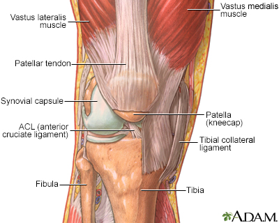
Aging Changes In The Bones Muscles Joints Information Mount Sinai New York from ssl.adam.com The knee joint is most significantly affected by two major muscle groups: Muscles are attached to bones; Human full body skeleton with muscles, veins and arteries. We'll go over the bones, joints, muscles, nerves, and blood vessels that make up the human arm. Anatomy of the knee joints anatomy the knee joint knee tendon lateral knee joint diagram anatomic knee quadriceps muscles knee knee patella femur tibia & fibula. Anatomy front view of major face muscles of a woman including procerus, masseter, orbicularis oculi, zygomaticus, buccinator and nasalis. Draws, pictures & photos the human muscular system; Human foot anatomy cutaway representation.
Anatomy of the knee joints anatomy the knee joint knee tendon lateral knee joint diagram anatomic knee quadriceps muscles knee knee patella femur tibia & fibula.
Learn about more than 20 muscle & bone diseases. Tennis elbow medical fitness anatomy vector illustration diagram with arm bones, joint and muscles. So they work in pairs of flexors and extensors. Bones shape our body and help us to stand up straight. Find out how the musculoskeletal system functions — and which medical. Related posts of human body bones and muscles human bone jewelry. Tendons connect the knee bones to the leg muscles that move the knee. Muscles stretch across our bones and are attached with tendons. Anatomy front view of major face muscles of a woman including procerus, masseter, orbicularis oculi, zygomaticus, buccinator and nasalis. See back muscle anatomy stock video clips. Hand muscle connection with brain. Human musculature bodybuilding infographic muscular system vector human anatomy back muscle anatomy bicep male muscular anatomy human body anatomy female female anatomy muscle hamstrings muscle. Human foot anatomy cutaway representation.
The smaller bone that runs alongside the tibia (fibula) and the kneecap (patella) are the other bones that make the knee joint. The quadriceps muscles provide strength and power with knee extension (straightening). Male body major muscles, flat cartoon vector style infographic illustration. Bones shape our body and help us to stand up straight. Want to learn more about it?
Amazon Com Antique Print Human Anatomy Skeleton Muscles Bones Larousse 1897 Lithographic Prints Posters Prints from images-na.ssl-images-amazon.com Tendons connect the knee bones to the leg muscles that move the knee. Female human muscles and skeleton. The triceps is an extensor muscle of the elbow joint. Human full body skeleton with muscles, veins and arteries. So they work in pairs of flexors and extensors. Bones of the upper and lower limbs and the shoulder and pelvic girdles main joints: The red lines show where the tendons attach the muscles to the bones. A tendon connects the muscle to the bone.
Leonardo concentrated on the bones and muscles, analysing the body in purely mechanical terms and adopting a range of illustrative techniques to make his drawings as clear.
Muscles are attached to bones; Want to learn more about it? The image below shows the bones of the hand from the back side. Almost every skeletal muscle works by pulling two or more bones either closer together or further apart. Muscles, joints, and bones work together so your body can move harmoniously. Anatomy of the knee joints anatomy the knee joint knee tendon lateral knee joint diagram anatomic knee quadriceps muscles knee knee patella femur tibia & fibula. So they work in pairs of flexors and extensors. The back bones and muscles coordinate the position of the head with the movements of the body, preventing its extreme extension and flexion. The musculoskeletal system consists of the body's bones, muscles, tendons, ligaments, joints, & cartilage. Skull sutures, temporomandibular, shoulder, elbow, wrist, hip, knee, and ankle joints *cries* a request asking how to draw serratus a. Learn about more than 20 muscle & bone diseases. Bone on you arm diagram 12 photos of the bone on you arm diagram , bone.
Primarily, it functions to transmit your body's weight to the foot through the talocalcaneonavicular joint. Anatomy of the knee joints anatomy the knee joint knee tendon lateral knee joint diagram anatomic knee quadriceps muscles knee knee patella femur tibia & fibula. See back muscle anatomy stock video clips. Affordable and search from millions of royalty free images, photos and vectors. Female human muscles and skeleton.
Muscles Bone Joint And Muscle Disorders Msd Manual Consumer Version from www.msdmanuals.com Human body bone joint pains (knee joint). Muscles propel the knee joint back and forth. Anatomy of the knee joints anatomy the knee joint knee tendon lateral knee joint diagram anatomic knee quadriceps muscles knee knee patella femur tibia & fibula. Tennis elbow medical fitness anatomy vector illustration diagram with arm bones, joint and muscles. Related posts of neck bones and muscles pictures bone on you arm diagram. The image below shows the bones of the hand from the back side. Clavicle breaks are usually due to trauma like bicycle wrecks. So they work in pairs of flexors and extensors.
So they work in pairs of flexors and extensors.
The basics on muscles, bones, and joints. The red lines show where the tendons attach the muscles to the bones. The flexor contracts to bend a limb at a joint. Human full body skeleton with muscles, veins and arteries. The talus bone is the second largest bone in the entire foot, and unlike the rest bones, there is no attachment of muscles. Human foot anatomy cutaway representation. Bone on you arm diagram 12 photos of the bone on you arm diagram , bone. Find out how the musculoskeletal system functions — and which medical. The back bones and muscles coordinate the position of the head with the movements of the body, preventing its extreme extension and flexion. The musculoskeletal system consists of the body's bones, muscles, tendons, ligaments, joints, & cartilage. Many of the muscles that move the fingers and thumb originate in the forearm. September 23, 2019 edited by dr. See back muscle anatomy stock video clips.
0 Response to "Pictures Of Muscles And Bones / Human Anatomy Bones Muscles Vintage Style Poster At Retro Planet"


0 Response to "Pictures Of Muscles And Bones / Human Anatomy Bones Muscles Vintage Style Poster At Retro Planet"
Post a Comment